


Excessive tension in the back muscles can cause a lot of discomfort and pain. Osteochondrosis destroys the structure of the vertebrae and intervertebral discs, causing severe compression of nerve endings. Usually, pathology is accompanied by deterioration of blood circulation, which causes the destruction of nutrients in the brain and internal organs.
Osteochondrosis-what?

Osteochondrosis is a recurrent disease that occurs in a chronic form and is accompanied by disc destruction of the vertebrae. Their organization is disturbed, resulting in a decrease in their elasticity and subsequent changes in shape. The intervertebral space gradually decreases. This leads to a loss of stability of the spine in the area of pathological development.
The process of pathological tissue destruction occurs in the background of pinched nerve endings, which are directly led out from the area where the spinal cord is located. As a result, the back muscles are in constant tension. In this case, patients will complain of back pain and other symptoms.
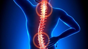
Based on the localized characteristics of the spine structure, these characteristics are covered by degenerative changes, so it is possible to distinguish the cervical spine, thoracic and lumbar s types in the pathological process. The main symptom of the development of osteochondrosis is pain, the intensity and severity of which usually increase during physical exercise.
There is stiffness in motion. In addition, the clinical image is characterized by the presence of signs of vertebral body type-headache, blood pressure changes, deterioration of visual function, hearing loss, etc.
Development mechanism
The development of osteochondrosis is related to the fact that the nucleus pulposus begins to lose its hydrophilicity. This semi-liquid structure contains connective tissue fibers and chondroitin (a gel-like substance). In the process of human development and growth, the process of reducing the vascular bed in the intervertebral disc is actively proceeding. Nutrition is provided in a dispersed manner, which is manifested as a spontaneous stability of concentration. This function makes it difficult to completely restore cartilage that has been damaged or the spine is under excessive pressure.
Due to the violation of hormonal background and human nutrition, pathological abnormalities become more obvious. Cartilage tissue begins to lack the nutrients needed for normal development. Therefore, the disease appears in the following form:
- Decrease in strength and elasticity;
- Change consistency parameters and configuration properties.
Against the background of flattening the intervertebral disc, a radial crack was formed in the annulus. As a result, the intervertebral distance decreases and the facet joints start to move. Over time, pathological changes encompass the types of connective tissue related to the annulus and ligaments.
As tissues are destroyed by the immune system, the amount of immunoglobulin produced increases. This stimulates the development of an aseptic inflammatory process, and edema is formed in the area where the facet joints are located. They also spread to adjacent soft tissues.
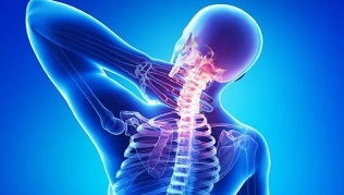
Due to the stretching of the joint capsule, the intervertebral disc loses its ability to fix the vertebrae. This instability in the position of the spinal structure increases the risk of nerve roots being crushed or blood vessels being crushed. This function is typical, for example, for cervical osteochondrosis, accompanied by strong oral symptoms.
Causes of diseases
Intervertebral disc disease will worsen as the tone of the spine skeletal muscle decreases. Due to the unreasonable and asymmetrical work of muscles, the destruction of cartilage tissue will occur with the long-term preservation of non-physiological positions of the human body. The violation was caused by wearing a heavy bag on the same shoulder, using a soft mattress and high pillows.
Due to a variety of internal and external negative factors, the destruction process of the intervertebral disc can be accelerated. These include:
- Abnormal endocrine mechanism and abnormal metabolism;
- Infectious pathology, including chronic;
- Injured the spine in the form of compression fractures and bruises;
- Normal body and prolonged hypothermia;
- Systemic and degenerative dystrophic diseases-gout, psoriasis, rheumatoid arthritis, osteoporosis, osteoarthritis;
- Smoking and alcohol abuse can destroy the state of the vascular system, impair blood circulation and cause cartilage to lack nutrients;
- Inadequate physical development, posture problems, flat feet-these defects increase the burden on the spine, because the amortization will not be enough;
- Obesity;
- Genetic susceptibility;
- Under normal pressure.
Symptoms

The main clinical symptom of any local (cervix, thoracic or lumbar s) osteochondrosis is pain syndrome. With recurrence, the pain begins to tingle and radiate to the vicinity of the body. Even if it moves slightly, it will increase. This forces the patient to place the torso in a forced position to minimize discomfort and soreness:
- suffering from osteochondrosis, it is best not to turn one head, but to turn the whole body;
- In patients with lumbar spine disease, when they sit down, stand upright, and move, they will have difficulty moving because they squeeze the nerves of the spine.
Usually, patients wake up in the morning and complain of dullness, constant pain and a feeling of stiffness. In this case, a differential diagnosis will be needed to help eliminate the risk of developing myositis due to inflammation of the skeletal spinal muscles or osteoarthritis.
Pain and tenderness are caused by compensatory tension in the muscle tissue. This condition is necessary to stabilize the spine movement area. Persistent mild or moderate pain will appear with the marked extension of the intervertebral disc and is caused by aseptic inflammatory changes.
A local feature of osteochondrosis is special symptoms:
- When suffering from cervical osteochondrosis, there will be pain in the neck area of the upper limbs. Head pain and numbness of the fingers were observed. If the disease manifests in a severe form, vertebral artery constriction may occur. In this case, patients began to complain about a marked deterioration in their health.
- The thoracic cavity is characterized by acute pain and pain in the back, and there is visceral pain syndrome in the heart area, right perichondrium and abdomen. The patient complained of numbness, abnormal skin sensations, shortness of breath, and crunching vertebrae.
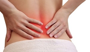
- Patients with lumbar osteochondrosis complain of pain in the back and lower limbs, and increase in strength when moving. Usually, it can be diagnosed with abnormal function of genitourinary system organs, male performance problems, and abnormal ovarian function. During the remission period, the pain may decrease. However, the influence of provocative factors led to its update.
- When mixed osteochondrosis is manifested, the symptomology can be manifested in multiple areas at the same time. This disease is characterized by a more serious course.
It should be remembered that vertebral displacement and osteophyte formation can cause compression of the vertebral artery. It provides nutrients to the brain and oxygen to its cells. When squeezing, food will be restricted, so patients will have problems with coordination, headaches, tinnitus and arterial hypertension.
Consequences of unhandled
The complicated course of osteochondrosis is due to the relatively rapid formation of hernia in the intervertebral disc. Their appearance is related to the displacement of the vertebral structure in the posterior direction. This will cause the longitudinal posterior ligament to rupture, resulting in an unstable position of the intervertebral disc, with various parts of the intervertebral disc extending into the spinal canal area. When the intervertebral disc with the nucleus pulposus penetrates into the root canal area, the hernia ruptures.
With the appearance of pathological abnormalities in the vertebral structure, the back of the brain began to squeeze, and the patient developed discogenic myelopathy. The symptoms of this condition are related to the numbness and weakness of certain muscle groups of the upper and lower limbs. Shows paresis, muscle atrophy and tendon reflexes. In some cases, there is a problem with emptying the bladder and bowel.
A herniated disc can squeeze the arteries that squeeze the spinal cord and is therefore very dangerous. The result of this pathology is the formation of an ischemic zone in which nerve cells have been damaged and died. The performance of nerve function is motor dysfunction, decreased tactile degree and nutritional disorders.
Disease diagnosis
Make a preliminary diagnosis based on the patient’s main complaint and symptoms. Experts study the condition of the spine in different positions, and recommend that the patient be at rest or in motion. In the next phase, the patient will be sent to a laboratory for diagnosis, which will help clarify or refute the diagnosis.
Research methods used include:
- Radiography-Provide a complete examination of the spine, and evaluate the condition of the vertebrae, diseases that exist in the form of growth and curvature. Experts will be able to determine the type of intervertebral interval, the state of the hole. In order to accurately identify osteochondrosis in the chest or cervical region, a two-stage X-ray examination is required. In the first stage, the patient lies beside him, and in the second stage, the patient lies directly on his back.
- The method of tomography by MRI or CTprovides very useful data to help study the vertebrae in detail without disturbing the organs covering the vertebrae. The picture shows the nerve and vascular system. MRI helps determine the signs of many spinal diseases and the location of injuries. Through CT, the hernia can be seen and the possible deviation of the spinal structure can be determined.
- Laboratory examinationTo evaluate the blood condition and its main parameters. Allows you to confirm the diagnosis and determine the possibility of developing accompanying diseases.
In many cases, as a result of the examination, the doctor diagnoses the existence of certain background diseases, which may be dangerous for its complications. For example, we are talking about hernia, herniation, radiculitis. Diagnosing the problem correctly can help effectively treat osteochondrosis. In this case, the disease itself is disguised as symptoms of other diseases in the early stages of development.
Treatment process
Osteochondrosis can be treated conservatively or surgically. The choice depends on the severity of the condition, the neglect of the condition, the degree of tissue degradation and the cause.
It is important to remember that it is impossible to completely cure osteochondrosis, because no medicine can completely restore the discs and vertebrae. The therapeutic effect is focused on inhibiting the destruction process and increasing the duration and stability of remission.
For symptomatic treatment, chondroprotective agents based on chondroitin sulfate or glucosamine are used.
Long-term testing has clinically confirmed the effectiveness of the treatment process with chondroprotective agents. If you use these funds for a long time within 3 months, the cartilage and other elements of the connection type (ligament-tendon instrument, bursa) will be partially restored.
The effectiveness of chondroprotective agents depends on the regularity of their intake. Otherwise, there will be no results. In the treatment of grade 3 osteochondrosis, ineffectiveness was also recorded, accompanied by severe cartilage destruction.
The following types of drugs can be used to relieve pain:
- Non-steroidal anti-inflammatory drugsHelp eliminate soft tissue inflammation caused by vertebral displacement. NSAID can effectively reduce pain, swelling and stiffness.
- Glucocorticoid methods-usually combined with anesthetics. They can relieve pain, restore immune mechanisms and provide anti-exudation effects.
- Muscle relaxants.They can effectively fight muscle spasms caused by nerves. They help relax skeletal muscles and have antispasmodic effects, preventing polysynaptic spinal reflexes.
- External therapy with warming effect.Special gels and ointments can stimulate receptors in subcutaneous tissues and activate blood flow. These drugs have analgesic and anti-edema effects.
With the help of medical equipment to activate blood flow, the vertebral body symptoms caused by pathological localization in the cervix or thoracic cavity can be eliminated. Nootropics and drugs to improve microcirculation are also prescribed. In some cases, you may need to take antidepressants and medications with anticonvulsants.
In the treatment of osteochondrosis, physical therapy is also used. UHF therapy, magnetic therapy, laser therapy, reflexology, massage, exercise therapy, hand and foot therapy, as well as swimming and yoga procedures can be prescribed. If conservative treatment fails, use microdiscectomy, puncture disc balancing, laser reconstruction, or implant replacement.