ankle arthritis

Ankle arthritis is a chronic disease that affects the articular cartilage and thus other structures of the joint (capsule, synovium, bones, ligaments). It is characterized by degenerative dystrophy. It presents with pain and limited movement, followed by a progressive impairment of support and walking functions. Diagnosis is made based on symptoms, examination, and radiography. Treatment is usually conservative with anti-inflammatory drugs, chondroprotectants, and corticosteroids, along with exercise therapy and physical therapy. In severe cases, hygienic arthroscopy, arthrodesis or endoprosthesis may be required.Ankle arthropathy

General information

Ankle arthritis is a disease in which joint cartilage and surrounding tissue are gradually destroyed. The disease is based on a degenerative dystrophic process; joint inflammation is secondary. Arthropathy has a chronic wave-like course, alternating with remission and exacerbation, and gradually progressing. Women and men suffer equally often. The potential for development increases dramatically with age. Meanwhile, experts say the disease is "getting younger" - with one in three cases of ankle arthritis now found in people under 45.

reason

Primary joints occur without any obvious cause. Secondary injuries to the ankle joint may occur under the influence of certain adverse factors. In both cases, the basis is metabolic processes that destroy cartilage tissue. The main causes and predisposing factors for the formation of secondary arthropathy of the ankle joint are:
  • Severe intra- and peri-articular injuries (talus fractures, ankle fractures, ligament tears and ruptures);
  • Ankle surgery;
  • Excessive load: excessive exercise intensity, walking for a long time, or standing frequently due to working conditions;
  • Wearing high heels, being overweight, and suffering from frequent microtrauma;
  • Diseases and conditions associated with metabolic disorders (diabetes, gout, pseudogout, postmenopausal estrogen deficiency);
  • Rheumatic diseases (systemic lupus erythematosus, rheumatoid arthritis);
  • Osteochondrosis of the lumbar spine, intervertebral hernias and other diseases associated with nerve compression and destruction of the musculature of the feet and legs.
Less commonly, causes of arthropathy are nonspecific septic arthritis, arthritis due to specific infections (tuberculosis, syphilis), and congenital developmental anomalies. Adverse environmental conditions and genetic predisposition play a role in the development of arthropathy.

onset

Typically, joint surfaces are smooth and elastic, slide smoothly relative to each other during movement, and provide effective shock absorption under load. Due to mechanical damage (trauma) or metabolic disorders, cartilage loses its smoothness and becomes rough and inelastic. Cartilage "rubbs" during movement and damages each other, leading to worsening of pathological changes.Due to insufficient depreciation, too much load is transferred to the underlying bone structure, and degenerative dystrophic diseases can also occur in them: the bones deform and grow along the edges of the joint area. Due to secondary trauma and disruption of the normal biomechanics of the joint, not only the cartilage and bone are damaged, but also the surrounding tissue.The joint capsule and synovium thicken, and fibrotic lesions form in the ligaments and periarticular muscles. The joint's ability to participate in movement and bear loads decreases. Instability develops and pain increases. In severe cases, the joint surface is destroyed, the supporting function of the limb is damaged, and movement is impossible.

symptom

Initially, after heavy loads, rapid fatigue and mild pain in the ankle joint are detected. Subsequently, the pain syndrome becomes more severe, changes in its nature and timing. The distinguishing features of joint pain are:
  • The pain begins. Appears after resting and then gradually disappears with exercise.
  • Dependence on load. The pain worsens during movement (standing, walking) and the joints tire quickly.
  • Pain at night. Usually appears in the morning.
The condition occurs in waves, with symptoms becoming more pronounced during exacerbations and first disappearing and then becoming less intense during remission phases. Symptoms may gradually progress over years or decades. In addition to pain, the following symptoms may occur:
  • There may be crunching, squeaking or clicking sounds when moving.
  • During an exacerbation of the condition, the area around the joint sometimes becomes swollen and red.
  • Because the joints are unstable, patients often twist their legs, resulting in ligament sprains and tears.
  • Stiffness and limited movement were noted.

complication

During exacerbations, reactive synovitis may occur, accompanied by intra-articular effusion. In later stages, significant deformation occurs. Movement is severely limited and contracture occurs. Support becomes difficult; when moving, the patient is forced to use crutches or a cane. Reduced or lost work ability.

diagnosis

The diagnosis of ankle arthritis is made by an orthopedic surgeon based on the results of investigations, external examination data, and additional studies. On initial examination, there may be no changes, but later deformation, limited movement, and pain on palpation may occur. The most important thing is visualization technology:
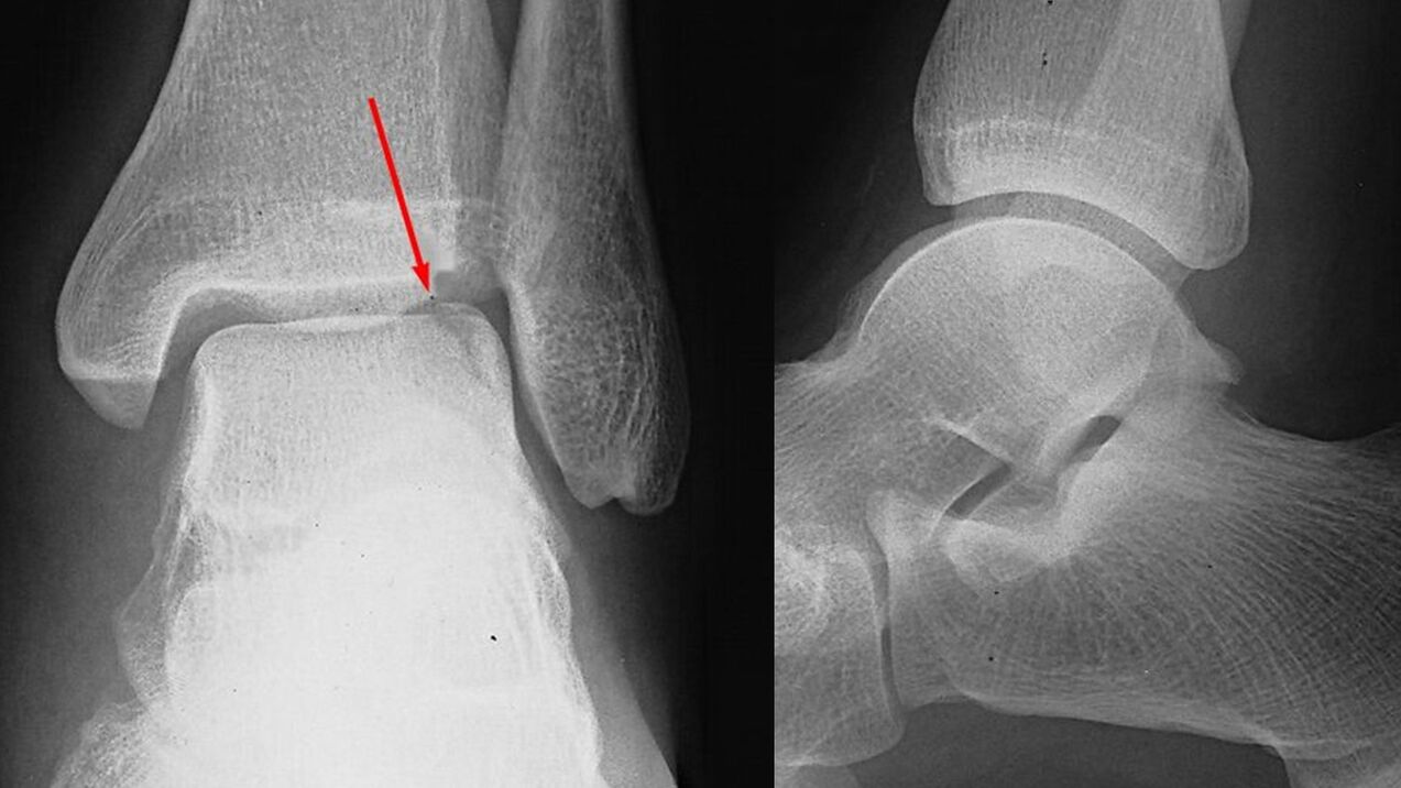
  • X-ray of the ankle joint. Plays a decisive role in making a diagnosis and determining the extent of joint disease. Pathological manifestations include narrowing of the joint space and hyperplasia (osteophytes) at the edges of the joint surface. In later stages, cystic formation and bone sclerosis are detected in the subchondral (located beneath the cartilage) areas of the bone.
  • Tomography studies. Use when directed. In difficult cases, in order to more accurately assess the condition of the bone structure, the patient also undergoes a computed tomography scan and examination of the soft tissues - an MRI of the ankle joint.
There are no changes to laboratory testing. If necessary, in order to determine the cause of the arthrosis and its differential diagnosis with other diseases, relevant specialists should be consulted: neurologist, rheumatologist, endocrinologist.Ankle X-ray

Ankle arthritis treatment

Treatment of the pathology is long-term and complex. Patients are typically seen on an outpatient basis by a plastic surgeon. During the exacerbation of the condition, admission to the trauma unit and orthopedics is possible. Lifestyle and correct physical activity patterns play the most important role in slowing down the progression of arthrosis, therefore, patients are advised to lose weight and optimize leg load.

medical treatement

It is selected individually, taking into account the stage of the arthrosis, the severity of symptoms and concomitant diseases. Including general agents and local agents. The following groups of medications are used:
  • General NSAIDs. Tablet form is usually used. Drugs have a certain negative impact on the gastric mucosa, so for gastrointestinal diseases, "mild" drugs should be chosen.
  • Local NSAIDs. Recommended for use during exacerbation and remission periods. Tablets can be used as an alternative if side effects occur. Available in ointment and gel forms.
  • Chondroprotectant. Substance that helps normalize the metabolic processes of cartilage tissue. They are used in the form of creams, gels, and preparations for intra-articular administration. Use medications containing glucosamine and collagen hydrolyzate.
  • Hormone agents. If medications do not relieve severe pain, intra-articular corticosteroid injections may be given no more than 4 times per year.
  • Metabolic stimulants. To improve local blood circulation and activate tissue metabolism, niacin is required.

Physiotherapy

Patients receive comprehensive physical therapy, which is tailored to the presentation and stage of the disease. The patient was referred for physical therapy. In the treatment of joint diseases, massage and ultrahigh frequencies are used. In addition, in pathological treatment they use:
  • Laser Treatment;
  • thermal program;
  • Drug electrophoresis and ultrasonic electrophoresis.

Surgery

It shows that in the late stage of the disease, when conservative treatment is ineffective, severe pain syndrome occurs, the patient's quality of life deteriorates, or the work ability is limited. These surgeries are performed in a hospital setting and are open and minimally invasive:
  • Arthroscopic intervention. If the cartilage is severely damaged, arthroscopic chondroplasty is performed. Hygiene arthroscopy (removal of structures that impede movement) is often used to treat severe pain in the second stage of arthropathy. The results last for years.
  • Arthrodesis of the ankle joint. It is performed with severe destruction of the joint surface and involves removal of the joint and "fusion" of the bones of the foot and lower leg. Restoring the support function of the limb in the event of loss of joint mobility.
  • Ankle endoprosthesis. Performed for advanced joint disease. Involves removing damaged joint surfaces of bones and replacing them with plastic, ceramic or metal prostheses. Activity is fully restored and the prosthesis has a service life of 20-25 years.

forecast

Changes in the joints are irreversible, but arthrosis progresses slowly, prompt initiation of treatment and compliance with the recommendations of an orthopedic traumatologist can, in most cases, preserve working ability and a high quality of life for decades after the first appearance. symptom. With the rapid increase in pathological changes, endoprostheses can prevent people from becoming disabled.

prevention

Preventative measures include reducing the severity of injuries, especially during winter ice periods. If you are obese, you should take measures to lose weight to reduce the load on your joints. You should maintain moderate physical activity, avoid overload and microtrauma, and promptly treat conditions that may cause ankle arthritis.